



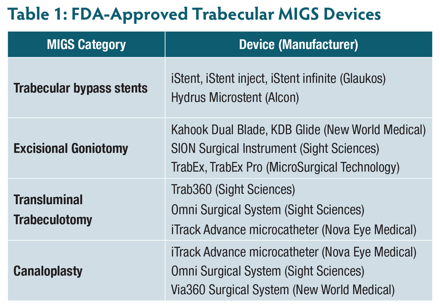
The landscape of glaucoma management has shifted substantially over the past decade. Minimally invasive glaucoma surgery (MIGS) has emerged as a preferred surgical option for patients with mild to moderate primary open-angle glaucoma (POAG), particularly those undergoing concurrent cataract extraction. Procedures that target the trabecular meshwork (TM) and Schlemm’s canal—including trabecular bypass stents, excisional goniotomy, transluminal trabeculotomy, and canaloplasty with or without goniotomy (Table 1)—lower intraocular pressure (IOP) and offer a favorable safety profile and rapid recovery.1,2
Yet MIGS, like all glaucoma interventions, is not curative. IOP can begin to rise as early as 12 to 24 months following surgery because of progressive TM dysfunction, stent fibrosis, malposition, changes in episcleral venous pressure, or distal collector channel resistance not addressed by the procedure. When IOP drifts above target, clinicians face a stepwise decision: escalate topical therapy, pursue laser treatment, or advance to more invasive filtration surgery.
Selective laser trabeculoplasty (SLT) occupies a potentially underappreciated position in this algorithm. Demonstrated in the landmark LiGHT trial to be noninferior to first-line topical therapy for untreated OAG, SLT lowers IOP by stimulating cytokine-mediated remodeling of the TM extracellular matrix, thereby enhancing aqueous outflow without causing coagulative trabecular scarring.3 At 6 years of follow-up, SLT proved superior to topical therapy: 70% of eyes in the SLT group required no additional medical or surgical treatment, disease progression occurred more frequently in the topical therapy group than in the SLT group (26.8% vs 19.6%; P=.006), and the need for surgical intervention was significantly reduced.4 Its repeatability, favorable adverse-effect profile, and office-based administration make it an attractive bridging intervention when MIGS efficacy wanes.
The Post-MIGS Status of the Trabecular Meshwork
MIGS procedures differ in how they affect the TM. Some preserve the TM and keep it largely intact, while others remove or bypass it across part or all of the angle. Because SLT depends on functional TM tissue to lower IOP, the degree of TM preservation after a given MIGS procedure directly determines whether SLT remains a viable subsequent option. This varies by procedure type—canal-based bypass stents, excisional goniotomy, transluminal trabeculotomy, or canaloplasty.
Bypass Stents
Trabecular bypass stents create direct conduits from the anterior chamber into Schlemm’s canal, reducing reliance on TM permeability without ablating or removing trabecular tissue. In this setting, the TM remains structurally intact in the quadrants surrounding the stent. SLT acts on pigmented TM cells through selective photothermolysis, upregulating metalloproteinases, stimulating macrophage-mediated phagocytosis of extracellular debris, and remodeling the juxtacanalicular matrix. These processes can theoretically occur in laser-naïve TM regardless of stent presence.
A retrospective study by Sieminski et al evaluated SLT following combined phacoemulsification and iStent implantation in eyes with inadequately controlled POAG. The investigators found a statistically significant IOP reduction of approximately 2 mmHg to 3 mmHg at 12 months, with medication reduction broadly comparable to that achieved with standalone SLT in a matched cohort. Notably, the presence of a stent did not appear to impair—and may even have modestly augmented—aqueous drainage, suggesting that SLT-driven outflow enhancement and stent-mediated bypass act through complementary rather than competing mechanisms.5
Excisional Goniotomy
Excisional goniotomy—for example with the Kahook Dual Blade (KDB)—removes the inner and outer walls of the TM over a 90° to 120° arc, creating a direct communication between the anterior chamber and Schlemm’s canal. Thus, in treated quadrants, the typical cellular substrate for SLT’s photobiologic effect is absent. However, 240° to 270° of TM typically remains, and these untreated regions may retain meaningful responsiveness to SLT.
A study by Dorairaj et al examining the relationship between prior SLT response and subsequent phaco/KDB outcomes found that eyes responding well to SLT before goniotomy also tended to achieve more robust postoperative IOP control, suggesting a shared distal outflow effector pathway.6
Transluminal Trabeculotomy
Gonioscopy-assisted transluminal trabeculotomy (GATT) unroofs Schlemm’s canal over 360°, effectively eliminating the entire circumferential TM as an outflow barrier.7 In this setting, the rationale for SLT is weakest because the primary target tissue has been removed throughout the angle. However, the distal outflow system—including the collector channels, aqueous veins, and episcleral venous bed—remains intact and may itself contribute to persistent outflow resistance.
Some investigators have proposed that SLT’s cytokine-mediated signaling may exert secondary effects beyond the immediate TM, but this remains speculative, and clinical evidence supporting SLT after 360° trabeculotomy is sparse. In practice, most eyes that fail GATT will likely require incisional surgery rather than repeat laser treatment.
Canaloplasty
Whether performed ab externo with a suture-tensioning technique or ab interno using viscodilation of Schlemm’s canal and the collector channel ostia (such as with iTrack or Via360), canaloplasty works by dilating the distal outflow system rather than excising or bypassing the TM. Importantly, the TM remains intact, making canaloplasty perhaps the most favorable MIGS category for preserving SLT target tissue.
The theoretical rationale for SLT after canaloplasty is therefore strong. If IOP rises following canal dilation because of progressive TM dysfunction or renarrowing of the canal, SLT-mediated cytokine remodeling of the TM could address the proximal resistance component that canaloplasty does not primarily target.
Published clinical data on SLT after canaloplasty are lacking, but the preserved angle anatomy and TM pigmentation suggest that standard SLT technique and laser settings need not be modified. Eyes that have undergone isolated canaloplasty without a concurrent goniotomy may represent some of the strongest candidates for post-MIGS SLT when IOP begins to rise above target.
Evidence for SLT After MIGS
Prospective data evaluating SLT after MIGS remain limited, but several retrospective studies and mechanistic analyses have begun to inform clinical practice.
Sieminski et al provided the most direct evidence for SLT after bypass-stent MIGS, demonstrating a clinically meaningful IOP reduction in eyes with prior iStent implantation and suboptimal pressure control. The mean IOP reduction was sustained through 12 months, and no adverse events specific to SLT in the post-MIGS setting were reported.5
Pahlitzsch et al examined the influence of prior SLT on subsequent iStent inject outcomes and found that patients who underwent SLT before MIGS achieved IOP reductions comparable to those without prior laser treatment, although the treatment effect trended modestly lower in heavily lasered angles. This finding raises the symmetric concern of whether residual TM responsiveness may likewise influence the effectiveness of SLT after MIGS.8
The most analogous data come from Lommatzsch et al, who evaluated SLT after failed phacoemulsification combined with excimer laser trabeculotomy (phaco-ELT), a canal-opening procedure with mechanistic similarities to MIGS trabeculotomy. Among 23 eyes, the median time to SLT failure was 7.2 months—shorter than typically observed in treatment-naïve eyes—but the procedure nonetheless delayed the need for incisional filtration surgery. The investigators concluded that SLT may serve as a useful temporizing measure, particularly for patients wishing to defer bleb-forming surgery.9
Taken together, the available evidence suggests that SLT is most likely to be effective after trabecular meshwork–preserving MIGS procedures, such as bypass stenting, partial goniotomy, and canaloplasty, and least likely to be effective after circumferential trabeculotomy.5,8,9 Although the duration of response may be shorter than in treatment-naïve eyes, SLT may still provide a clinically meaningful opportunity to delay more invasive surgery, and patients should be counseled accordingly.
Patient Selection for Post-MIGS SLT
Identifying appropriate candidates for SLT after MIGS requires consideration of the prior procedure, gonioscopic anatomy, glaucoma severity, and patient preference.
Preserved TM Anatomy
Gonioscopy should be performed before SLT to confirm that visible, pigmented TM remains in the quadrants to be treated. Bypass stent procedures (iStent inject, Hydrus) generally preserve TM throughout the angle, whereas partial goniotomy typically leaves 240° to 270° of intact TM accessible to laser treatment. By contrast, 360° trabeculotomy leaves little meaningful TM target and is generally not a favorable substrate for SLT.
Mild to Moderate Disease With Adequate Optic Nerve Reserve
The IOP-lowering effect of SLT—typically 20% to 30% from an elevated baseline—may be insufficient to achieve target IOP in advanced disease requiring substantial pressure reduction. In patients with cup-to-disc ratios approaching 0.9 and markedly constricted visual fields, the risk of progressive damage during a trial of SLT may outweigh the potential benefit of delaying filtration surgery.
Elevated IOP
Consistent with the primary SLT literature—including the predictive factor analysis by Chen et al, which identified a baseline IOP greater than 18 mmHg as the strongest predictor of treatment success10—post-MIGS SLT is most likely to be effective when there is meaningful IOP elevation to correct. Eyes that have drifted only modestly above target (eg, from 13 mmHg to 15 mmHg) may experience a smaller absolute response than eyes with a more substantial increase in IOP.
No Active Inflammation or Significant PAS
Postoperative inflammation or peripheral anterior synechiae (PAS) related to prior MIGS can obscure the angle, interfere with laser delivery, and impair TM cellularity. These conditions should be resolved before SLT is attempted.
When to Consider SLT After MIGS
No consensus guideline specifies the optimal interval between MIGS and subsequent SLT, but several practical considerations can help guide timing.
Most clinicians would defer SLT for at least 3 months after MIGS to allow postoperative inflammation and hyphema to resolve and IOP to stabilize. During this period, transient IOP elevation may reflect postoperative inflammation or a steroid response rather than true MIGS failure, and premature laser treatment could confound assessment.
Beyond 3 months, the decision to proceed with SLT should be guided by evidence that IOP has consistently exceeded target on 2 or more visits, that medication adjustment alone is insufficient or poorly tolerated, and that gonioscopy confirms the presence of treatable TM. Some surgeons prefer to wait at least 6 months to ensure that the full effect of MIGS has been realized and is beginning to wane rather than continuing to evolve.
When the TM is accessible throughout the angle, a practical approach is to perform 360° SLT. In eyes that have undergone partial goniotomy, treatment can be directed to the remaining TM. Postlaser IOP monitoring is based on surgeon preference, although patients are typically reassessed at 6 to 8 weeks to evaluate treatment response. If IOP has not returned to target within 3 months, the treatment algorithm should be reassessed, and filtration surgery should be considered.
The comparison by Klamann et al of SLT and MIGS as sequential vs parallel interventions supports a stepwise escalation strategy, positioning SLT as a less tissue-disruptive option before filtration surgery when angle anatomy permits.11
SLT in the Post-MIGS Treatment Algorithm
The interventional glaucoma paradigm—SLT first, followed by MIGS at the time of cataract surgery, with subsequent escalation as needed—has been endorsed by expert consensus statements.1 In current practice, SLT often precedes MIGS. However, a growing cohort of patients underwent MIGS as their initial intervention, particularly those who had cataract surgery before the widespread adoption of SLT as first-line therapy. In these patients, the role of SLT after MIGS represents a clinically distinct and underrecognized management question.
For eyes with an IOP rise after MIGS, the following algorithm may be useful:
- Confirm stent or device patency and position by gonioscopy, and rule out malposition, obstruction, or PAS.
- Assess medication adherence.
- If viable TM is present in at least 180° of the angle, consider SLT as the next procedural step,5,9 particularly in patients with mild to moderate disease, IOP above target, and a preference to avoid additional medications or more invasive surgery.
- Counsel patients that the duration of response after post-MIGS SLT may be shorter than that observed in treatment-naïve eyes.
- If SLT fails or TM anatomy is unfavorable (eg, after 360° trabeculotomy), proceed to subconjunctival or supra- choroidal drainage procedures based on disease severity and surgeon preference.
This stepwise approach reserves bleb-forming surgery for when it is truly needed, minimizes cumulative medication burden, and respects patient preferences. It is consistent with the broader interventional glaucoma philosophy of exhausting less invasive options before proceeding to procedures associated with greater risk and recovery.1
Conclusion
SLT retains a meaningful role in the management of IOP elevation following trabecular- and canal-based MIGS, provided the clinician carefully assesses gonioscopic anatomy, disease severity, and the type of prior procedure. Eyes with preserved TM after canaloplasty, bypass stenting, or partial goniotomy are the best candidates, and these patients can reasonably expect a clinically meaningful IOP reduction that may delay the need for incisional surgery.5,9,11 By contrast, eyes that have undergone circumferential trabeculotomy are generally poor candidates for SLT, and escalation to other surgical options may be warranted.7 As the population of patients undergoing MIGS continues to grow, so too will the need for clear algorithms to guide subsequent management, including the appropriate role of SLT.
Prospective, controlled studies evaluating SLT outcomes according to MIGS procedure type and postoperative angle anatomy are needed to refine these recommendations. Until such data are available, gonioscopy-guided clinical judgment, informed by the mechanistic rationale and early retrospective evidence summarized here, provides a reasonable framework for incorporating SLT into the post-MIGS treatment paradigm.1,3,4 GP

References
1. Bacharach J, Reda R, Alfaro A, et al. Interventional glaucoma consensus treatment protocol. Clin Ophthalmol. 2025;19:497-519. doi:10.2147/OPTH.S500382
2. Lusthaus J, Goldberg I. Current management of glaucoma. Med J Aust. 2019;210(4):180-187. doi:10.5694/mja2.50020
3. Gazzard G, Konstantakopoulou E, Garway-Heath D, et al; LiGHT Trial Study Group. Selective laser trabeculoplasty versus eye drops for first-line treatment of ocular hypertension and glaucoma (LiGHT): a multicentre randomised controlled trial. Lancet. 2019;393(10180):1505-1516. doi:10.1016/S0140-6736(18)32213-X
4. Gazzard G, Konstantakopoulou E, Garway-Heath D, et al; LiGHT Trial Study Group. LiGHT Trial: 6-year results of primary selective laser trabeculoplasty versus eye drops for the treatment of glaucoma and ocular hypertension. Ophthalmology. 2023;130(2):139-151. doi:10.1016/j.ophtha.2022.08.009
5. Sieminski SF, Bhatt P, Chen H, Davila JR, Bhatt A, Danias J. Efficacy of selective laser trabeculoplasty after iStent implantation in primary open-angle glaucoma. J Pers Med. 2021;11(8):797. doi:10.3390/jpm11080797
6. Dorairaj SK, Bhattacharya S, Williamson BK, et al. Selective laser trabeculoplasty and outcomes of subsequent phacoemulsification combined with Kahook Dual Blade goniotomy. Ophthalmol Ther. 2022;11(5):1767-1780. doi:10.1007/s40123-022-00554-3
7. Grover DS, Godfrey DG, Smith O, Feuer WJ, Montes de Oca I, Fellman RL. Gonioscopy-assisted transluminal trabeculotomy, ab interno trabeculotomy: technique report and preliminary results. Ophthalmology. 2014;121(4):855-861. doi:10.1016/j.ophtha.2013.11.001
8. Pahlitzsch M, Torun N, Gonnermann J, Maier AKB, Bertelmann E, Klamann MKJ. Influence of selective laser trabeculoplasty (SLT) on the iStent inject outcomes. Graefes Arch Clin Exp Ophthalmol. 2020;258(12):2773-2779. doi:10.1007/s00417-020-04887-6
9. Lommatzsch C, Vossmerbaeumer U, Jehle T, Bertram B, Heinz C. Selective laser trabeculoplasty following failed combined phacoemulsification cataract extraction and excimer laser trabeculotomy can control intraocular pressure for a limited time. Graefes Arch Clin Exp Ophthalmol. 2022;260(6):1979-1986. doi:10.1007/s00417-021-05428-x
10. Chen E, Golubchenko A, Javanbakht A, Fagerholm P. Predictive factors for outcomes of selective laser trabeculoplasty. J Glaucoma. 2020;29(6):468-473. doi:10.1097/IJG.0000000000001493
11. Klamann MKJ, Maier AKB, Gonnermann J, et al. Selective laser trabeculoplasty versus MIGS: forgotten art or first-step procedure in selected patients with open-angle glaucoma. Ophthalmol Ther. 2021;10(3):577-590. doi:10.1007/s40123-021-00347-0