



Angle-based minimally invasive glaucoma surgery (MIGS) has become an increasingly important option for patients with open-angle glaucoma. As these procedures are adopted more widely and often earlier in the disease course, the challenge for clinicians is identifying the patients most likely to benefit from angle surgery. The answer remains unclear because patient outcomes vary, demonstrating the complexity of the conventional outflow pathway.
One of the clearest examples of mechanism-based surgical selection is seen in MYOC-associated juvenile open-angle glaucoma (JOAG). Boese et al reported 4 children with MYOC-associated glaucoma who underwent 360° gonioscopy-assisted transluminal trabeculotomy (GATT). All 8 eyes achieved marked and durable intraocular pressure (IOP) reduction, discontinued glaucoma medications, and maintained stable vision over years of follow-up.1 Because MYOC mutations primarily affect the trabecular meshwork, GATT directly targets the presumed site of disease. The authors described this approach as genetically directed glaucoma surgery.
This concept is compelling but remains in the early stages. Current evidence remains limited to small case series and observational studies. At present, genetic testing should be viewed as an emerging precision medicine tool rather than a validated clinical predictor of the success of angle surgery.
Large registry studies provide a broader real-world view of outcomes of angle surgery. Yang et al analyzed 79,363 eyes undergoing MIGS in the Intelligent Research in Sight (IRIS) Registry from 2013 to 2019 and found that outcomes varied by procedure type, baseline characteristics, and whether surgery was performed alone or combined with phacoemulsification.2 These findings suggest that no single preoperative factor can reliably predict success in every patient.
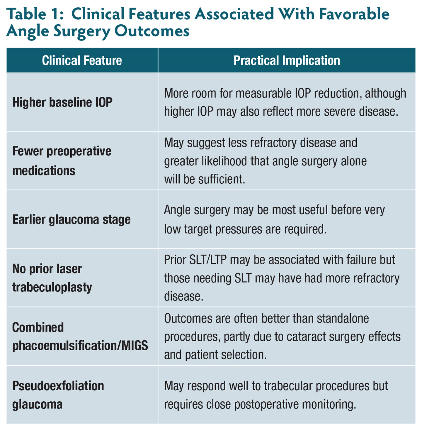
Still, several clinical features are consistently associated with more favorable outcomes (Table 1). These features can help frame preoperative counseling and guide procedure selection.
Baseline IOP: More Room to Lower Pressure
Baseline IOP is one of the most consistent clinical factors associated with postoperative response. Chan et al found that each 1 mmHg increase in preoperative IOP was associated with an 8% increase in the likelihood of surgical success after combined phacoemulsification/MIGS.3
However, this relationship should be interpreted carefully. Higher baseline IOP can be associated with greater absolute IOP reduction, but it may also reflect more severe or difficult-to-control glaucoma. Yang et al similarly found that eyes with higher baseline IOP tended to achieve greater pressure reduction, while higher baseline IOP was also associated with a greater risk of reoperation in some settings.2
In practice, a patient with a higher baseline IOP may experience a larger numerical drop, but that does not necessarily mean the eye is at lower risk. The target IOP, disease severity, and likelihood of needing future intervention should still guide counseling.
Medication Burden and Disease Stage
Medication burden is another useful clinical clue. Eyes requiring fewer preoperative glaucoma medications may be more likely to achieve medication-free success. Paletta Guedes et al identified a lower medication burden as a factor associated with unqualified success after trabecular bypass surgery.4 On the other hand, Chan et al found that a higher baseline number of IOP-lowering medications was associated with a greater likelihood of success, which in that study included medication reduction.3 Therefore, medication burden should be interpreted alongside washout IOP, which may better reflect the eye’s untreated pressure and outflow reserve.
Disease stage also matters. Earlier-stage glaucoma has been associated with better outcomes, whereas moderate-to-severe glaucoma has been associated with a higher risk of reoperation in registry analyses.2,4 Clinically, this fits with the role of angle surgery as a procedure that often provides moderate IOP lowering. In advanced glaucoma, where very low target pressures may be needed, conventional outflow enhancement may be insufficient as a standalone strategy.
Combined Surgery vs Standalone MIGS
Whether angle surgery is performed with cataract surgery is another important consideration. Outcomes are often more favorable when MIGS is combined with phacoemulsification. Paletta Guedes et al found that combined phacoemulsification/MIGS was associated with surgical success, and Yang et al reported substantially lower reoperation rates for combined procedures than for standalone procedures.2,4
This difference likely reflects several factors. Cataract surgery itself can lower IOP.5 In addition, patients undergoing standalone MIGS may have more refractory disease or may already be pseudophakic, limiting the additional benefit of lens extraction. Therefore, it would be helpful to distinguish between “MIGS at the time of cataract surgery” and “standalone MIGS for glaucoma control” when counseling patients.
Prior SLT: A Useful Clinical Clue
Prior selective laser trabeculoplasty (SLT) may be a useful historical factor when evaluating angle surgery. Chan et al found that prior SLT was negatively associated with success after combined phacoemulsification/MIGS.3 Mitchell et al later analyzed 164,965 angle-based MIGS procedures in the IRIS Registry and found that prior laser trabeculoplasty was associated with increased risk of subsequent glaucoma reoperation, with an adjusted hazard ratio of 1.53.6
However, this association should not be interpreted definitively as proof that SLT causes worse MIGS outcomes. More likely, prior SLT may identify eyes with more treatment-resistant conventional outflow disease.
In clinical practice, a history of SLT can serve as a useful counseling point. It does not preclude angle surgery but may warrant more cautious expectations, particularly if the response to SLT was poor or short lived.
Glaucoma Subtype: Why Pseudoexfoliation Matters
The glaucoma subtype may also influence surgical response. Pseudoexfoliation glaucoma (PXG) is often considered to be more aggressive than primary open-angle glaucoma (POAG), but it may also be particularly responsive to procedures that target the trabecular meshwork because pseudoexfoliative material contributes to resistance at or near the trabecular outflow pathway.
Aktas et al compared Prolene GATT outcomes in PXG and POAG. At 1 year, PXG eyes achieved greater IOP reduction than POAG eyes, although the differences diminished over time, and success rates were similar by 3 years.7 Bektas et al also reported favorable 5-year outcomes after GATT in PXG, with qualified success rates of 91.2% for PXG compared with 83.1% for POAG.8
At the same time, PXG remains clinically challenging. Fujita et al found that pseudoexfoliation patients were more likely to undergo glaucoma surgery overall and experienced more postoperative IOP spikes and additional surgeries compared with patients with POAG.9 Taken together, these studies suggest that PXG may respond particularly well to trabecular procedures, although these patients require close postoperative monitoring.
Imaging Biomarkers: Moving Beyond Clinical Clues
Traditional clinical variables are helpful but may offer limited guidance in surgical selection. Increasingly, investigators are exploring whether imaging biomarkers can better characterize the conventional outflow pathway and predict surgical response. Anterior-segment optical coherence tomography angiography (OCTA) is one promising approach. Okamoto et al found that eyes with lower preoperative deep, or intrascleral, vessel density had higher surgical success rates after trabecular meshwork-targeted MIGS.10 One possible explanation is that a lower baseline intrascleral vascular signal may indicate greater capacity for postoperative outflow recruitment.
Trabecular meshwork thickness may also be useful. Yoon et al found that greater preoperative trabecular meshwork thickness was associated with iStent inject W (Glaukos) implantation failure.11 Interestingly, conventional angle parameters such as angle opening distance and trabecular-iris angle were not predictive.
These findings challenge the assumption that an open angle alone is enough to predict MIGS success. At the current stage, imaging biomarkers remain an active area of investigation. Existing studies are small and require prospective validation. Nonetheless, they suggest that MIGS success depends not only on gross angle anatomy but also on the functional status of the conventional outflow pathway.
The Distal Outflow Problem
Even with ideal patient selection, angle surgery has inherent physiologic limits. Most angle-based procedures target the trabecular meshwork or Schlemm’s canal, but aqueous outflow depends on the entire conventional pathway. If resistance is predominantly distal, trabecular bypass or incision alone may have a limited effect.
Dickerson and Brown reviewed the concept of segmental aqueous outflow, emphasizing that not all collector channels are equally active.12 Aqueous angiography studies have shown that outflow is segmental, meaning that some regions of the angle contribute more to aqueous drainage than others.13,14 A trabecular bypass placed near active collector channels may therefore perform differently from a stent placed in a relatively inactive region. A Cochrane overview and network meta-analysis by Bicket et al also found that Hydrus Microstent (Alcon) provided additional IOP lowering compared with phacoemulsification alone and increased the likelihood of medication-free status.15 These findings suggest that procedure selection may matter, because treatments that address a broader segment of Schlemm’s canal or the distal outflow pathway may offer advantages in selected patients.
Limitations of the Evidence
Despite growing interest in predicting outcomes of angle surgery, the evidence base has important limitations. Many studies are retrospective and single-center, which introduces selection bias and limits generalizability.2,3,7,8 Surgeons often choose procedures based on a variety of factors, making it difficult to isolate the effect of any single variable.
Registry studies offer large sample sizes but have limitations as well. In IRIS Registry analyses, reoperation is often used as a surrogate for surgical failure because medication data may be incomplete.2,6,9 This approach likely underestimates the true failure rate, because many patients who do not reach the target IOP are managed with additional medications rather than repeat surgery.
Imaging biomarker studies are promising but remain small in scale. The anterior-segment OCTA study by Okamoto et al included 37 patients, and the trabecular meshwork thickness study by Yoon et al included 28 patients.10,11 These findings therefore require prospective, multicenter validation before they can be incorporated into clinical decision-making.
Finally, many MIGS studies include procedures performed in conjunction with cataract surgery, making it difficult to separate the effect of the angle procedure from phacoemulsification. This distinction is critical when counseling patients about standalone MIGS.
Conclusion: Toward Better Patient Selection
As angle-based MIGS procedures are adopted earlier and more broadly, identifying the patients most likely to benefit has become an important clinical priority. Current evidence suggests that favorable outcomes are more commonly observed in eyes with higher baseline IOP, earlier glaucoma stage, no prior laser trabeculoplasty, and procedures performed in combination with phacoemulsification.2-4,6 Additionally, glaucoma subtypes require careful consideration. Specifically, PXG may respond well to trabecular procedures, but these patients require careful postoperative monitoring because of their more aggressive disease course.7-9
Further studies on imaging and physiologic biomarkers may offer promising predictions on surgical success. Anterior-segment OCTA, trabecular meshwork thickness, aqueous angiography, and other methods for assessing distal outflow could help identify patients in whom the conventional outflow pathway can be successfully recruited. Genetic and molecular markers may eventually add another layer of precision, particularly in childhood and juvenile glaucoma. These tools may eventually allow clinicians to move beyond broad clinical categories and toward individualized procedure selection.
However, substantial gaps remain. Progress will require prospective, multicenter studies with standardized outcome definitions, longer follow-up, diverse patient populations, and a clear distinction between standalone and combined procedures. These studies should integrate clinical characteristics with imaging biomarkers and, potentially, genetic or molecular markers to develop more reliable models of surgical response. As our understanding of the conventional outflow pathway evolves, predicting success in angle surgery may shift from a practice rooted in clinical intuition to a precise, evidence-based approach. GP


References
1. Boese EA, Alward WLM, Fingert JH. Gonioscopy-assisted transluminal trabeculotomy for myocilin juvenile glaucoma. Ophthalmol Glaucoma. 2022;5(3):369-370. doi:10.1016/j.ogla.2021.07.003
2. Yang SA, Ciociola EC, Mitchell W, et al. Effectiveness of microinvasive glaucoma surgery in the United States: Intelligent Research in Sight Registry Analysis 2013-2019. Ophthalmology. 2023;130(3):242-255. doi:10.1016/j.ophtha.2022.10.021
3. Chan W, Zhang C, Mittal A, Fink A, Michalovic S, Weiner A. Effect of preoperative trabecular meshwork pigmentation and other eye characteristics on outcomes of combined phacoemulsification/minimally invasive glaucoma surgery. Ophthalmol Glaucoma. 2024;7(3):271-281. doi:10.1016/j.ogla.2024.01.001
4. Paletta Guedes RA, Gravina DM, Paletta Guedes VM, Chaoubah A. Factors associated with unqualified success after trabecular bypass surgery: a case-control study. J Glaucoma. 2020;29(11):1082-1087. doi:10.1097/IJG.0000000000001626
5. Hussein O, Musleh A, Abukhaled Y, Hammad A, Feras R, Abu Serhan H. Does standalone phacoemulsification lower intraocular pressure in glaucomatous eyes? A systematic review and meta-analysis. Eye (Lond). 2025;39(13):2518-2526. doi:10.1038/s41433-025-03927-7
6. Mitchell W, Yang SA, Ondeck C, et al. Effectiveness of angle-based minimally invasive glaucoma surgery after laser trabeculoplasty: an analysis of the IRIS(R) Registry (Intelligent Research in Sight). Ophthalmol Glaucoma. 2024;7(4):335-344. doi:10.1016/j.ogla.2024.03.003
7. Aktas Z, Ozdemir Zeydanli E, Uysal BS, Yigiter A. Outcomes of Prolene gonioscopy assisted transluminal trabeculotomy in primary open-angle glaucoma and pseudoexfoliation glaucoma: a comparative study. J Glaucoma. 2022;31(9):751-756. doi:10.1097/IJG.0000000000002063
8. Bektas C, Ozmen MC, Acar B, Uysal BS, Aktas Z. Long-term outcomes of gonioscopy-assisted transluminal trabeculotomy in open-angle glaucoma. J Glaucoma. 2026;35(2):92-99. doi:10.1097/IJG.0000000000002644
9. Fujita A, Elze T, Zhao Y, et al. Comparison of glaucoma surgery incidence and outcomes in pseudoexfoliation and primary open-angle glaucoma: IRIS(R) Registry analysis. Ophthalmology. 2026;133(2):214-222. doi:10.1016/j.ophtha.2025.09.012
10. Okamoto Y, Akagi T, Kameda T, et al. Prediction of trabecular meshwork-targeted micro-invasive glaucoma surgery outcomes using anterior segment OCT angiography. Sci Rep. 2021;11(1):17850. doi:10.1038/s41598-021-97290-8
11. Yoon H, Lee J, Lee SM, Lee JE, Kim SJ, Moon S. Trabecular meshwork thickness measured by swept-source AS-OCT as a predictor of surgical outcomes after trabecular micro-bypass stent implantation. J Clin Med. 2026;15(9):3341. doi:10.3390/jcm15093341
12. Dickerson JE, Jr., Brown RH. Circumferential canal surgery: a brief history. Curr Opin Ophthalmol. 2020;31(2):139-146. doi:10.1097/ICU.0000000000000639
13. Akagi T, Uji A, Huang AS, et al. Conjunctival and intrascleral vasculatures assessed using anterior segment optical coherence tomography angiography in normal eyes. Am J Ophthalmol. 2018;196:1-9. doi:10.1016/j.ajo.2018.08.009
14. Akagi T, Uji A, Okamoto Y, et al. Anterior segment optical coherence tomography angiography imaging of conjunctiva and intrasclera in treated primary open-angle glaucoma. Am J Ophthalmol. 2019;208:313-322. doi:10.1016/j.ajo.2019.05.008
15. Bicket AK, Le JT, Azuara-Blanco A, et al. Minimally invasive glaucoma surgical techniques for open-angle glaucoma: an overview of Cochrane Systematic Reviews and Network meta-analysis. JAMA Ophthalmol. 2021;139(9):983-989. doi:10.1001/jamaophthalmol.2021.2351