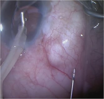
It is often thought that a large part of glaucoma surgery is the art of operating on and managing the conjunctiva. The success of trabeculectomy and subconjunctival bleb procedures, such as implantation of a Xen gel stent (Allergan), depends largely on a successful bleb. The bleb is an area that allows aqueous humor to flow out of the surgical site. The perfect bleb allows for a pathway for the aqueous to leave the anterior chamber through a natural passageway behind the conjunctiva, where it will be reabsorbed by surrounding tissue. The success of a bleb depends upon the surface area, as well as the vascular permeability. If it becomes scarred down at any point along this passageway, this will result in too much resistance to that flow. Lysing the scars around a scarred bleb allows for reinitiating flow into and out of the bleb, thereby facilitating reduction of intraocular pressure (IOP).
Blebs can come is different varieties. An ideally functioning bleb is one that exhibits diffuse, regular elevation with minimal vascularity, minimal thinning, and few cystic features. Flat blebs indicate diffuse scarring and a complete lack of flow. An encapsulated bleb demonstrates a classic “ring of steel,” a semicircular area of scarring in the conjunctiva that resists all flow from the anterior chamber, thereby causing an increase in pressure (Figure 1).

Bleb Needling
To lyse the scarred Tenon's to the conjunctiva, one can perform bleb needling to revise the bleb. There are multiple ways to perform bleb needling. It is most commonly thought of as an in-office procedure performed at the slit lamp, but it can also be done intraoperatively.1-7
Intraoperative Bleb Needling
Intraoperative needling is my preferred method of bleb needling. Although it is less commonly done because it requires a trip to the operating room, there are a number of reasons a surgeon might prefer it. First, it allows for the patient to be relaxed and supine. Second, it allows for the surgeon to use their dominant hand and a microscope. Third, it allows the surgeon to use an infusion into the anterior chamber so that there is constant flow, and one can visualize the successful needling and breaking through the scarred bleb and successfully achieving outflow of the aqueous into the subconjunctival space (Video 1).
Steps and tips for intraoperative bleb needling are as follows:
- Place betadine and topical lidocaine jelly.
- Make a temporal main wound with a keratome and use either a Lewicky cannula or the irrigation and aspiration handpiece to create an infusion into the eye.
- Use a bent 25-gauge needle on a tuberculin (TB) syringe to enter the conjunctiva superotemporally to the scarred area and use a side-to-side sweeping motion to lyse the scar tissue using the edge of the bevel until you see a raised bleb. Having the constant infusion enables the surgeon to see the bleb rise and confirms successful outflow.
- Inject 5-fluorouracil (5FU) or mitomycin into the subconjunctiva to help prevent further encapsulation and scar tissue formation.
In-office Bleb Needling
Tips for in-office bleb needling are similar to those described above, except that infusion is not placed. Instead, a speculum is placed, the eye is everted down with a cotton-tip swab in the nondominant hand, and a 25-gauge bent needle on a TB syringe is used to enter the conjunctiva and lyse the scar tissue while the patient is sitting upright at the slit lamp. The TB syringe can contain 5FU so that convenient administration using the same needle and track can be performed. The advantage of performing this in the office primarily is avoiding a trip to the operating room.
Adjunctive antimetabolites are commonly placed under the conjunctiva to help reduce scarring. The most commonly used antifibrotic agent placed in the office is 5FU. However, mitomycin, triamcinolone, and bevacizumab (Avastin; Genentech) have also been used.8-10 Postoperative care includes using a topical antibiotic following the procedure.
Conclusion
Bleb needling offers a method of reviving a bleb. If done correctly, bleb needling can reduce the IOP and avoid more invasive procedures, such as revision of a trabeculectomy or alternate glaucoma procedures. GP
References
- Ferrer H. Conjunctival dialysis in the treatment of glaucoma recurrent after sclerectomy. Am J Ophthalmol. 1941;24:788-790.
- Pederson JE, Smith SG. Surgical management of encapsulated filtering blebs. Ophthalmology. 1985;92(7):955-958. doi:10.1016/s0161-6420(85)33949-0
- Ewing RH, Stamper RL. Needle revision with and without 5-fluorouracil for the treatment of failed filtering blebs. Am J Ophthalmol. 1990;110(3):254-259. doi:10.1016/s0002-9394(14)76340-8
- Shin DH, Juzych MS, Khatana AK, Swendris RP, Parrow KA. Needling revision of failed filtering blebs with adjunctive 5-fluorouracil. Ophthalmic Surg. 1993;24(4):242-248. doi:10.3928/1542-8877-19930401-05
- Mardelli PG, Lederer CM Jr, Murray PL, Pastor SA, Hassanein KM. Slit-lamp needle revision of failed filtering blebs using mitomycin C. Ophthalmology. 1996;103(11):1946-1955. doi:10.1016/s0161-6420(96)30403-x
- Shin DH, Kim YY, Ginde SY, et al. Risk factors for failure of 5-fluorouracil needling revision for failed conjunctival filtration blebs. Am J Ophthalmol. 2001;132(6):875-880. doi:10.1016/s0002-9394(01)01232-6
- Broadway DC, Bloom PA, Bunce C, Thiagarajan M, Khaw PT. Needle revision of failing and failed trabeculectomy blebs with adjunctive 5-fluorouracil: survival analysis [published correction appears in Ophthalmology. 2005 Jan;112(1):66]. Ophthalmology. 2004;111(4):665-673. doi:10.1016/j.ophtha.2003.07.009
- Gutiérrez-Ortiz C, Cabarga C, Teus MA. Prospective evaluation of preoperative factors associated with successful mitomycin C needling of failed filtration blebs. J Glaucoma. 2006;15(2):98-102. doi:10.1097/00061198-200604000-00004
- Tham CC, Li FC, Leung DY, et al. Intrableb triamcinolone acetonide injection after bleb-forming filtration surgery (trabeculectomy, phacotrabeculectomy, and trabeculectomy revision by needling): a pilot study. Eye (Lond). 2006;20(12):1484-1486. doi:10.1038/sj.eye.6702372
- Kahook MY, Schuman JS, Noecker RJ. Needle bleb revision of encapsulated filtering bleb with bevacizumab. Ophthalmic Surg Lasers Imaging. 2006;37(2):148-150. doi:10.3928/1542-8877-20060301-12


